Home
/ Plant Cell Images Under Microscope : Plant Cells Under Microscope Stock Photo By C Solstudio 103192844 : A scanning electron microscope (sem) is a type of electron microscope that produces images of a sample by scanning the surface with a focused beam of electrons.
Plant Cell Images Under Microscope : Plant Cells Under Microscope Stock Photo By C Solstudio 103192844 : A scanning electron microscope (sem) is a type of electron microscope that produces images of a sample by scanning the surface with a focused beam of electrons.
Plant Cell Images Under Microscope : Plant Cells Under Microscope Stock Photo By C Solstudio 103192844 : A scanning electron microscope (sem) is a type of electron microscope that produces images of a sample by scanning the surface with a focused beam of electrons.. Browse 153 plant cells under microscope stock photos and images available, or start a new search to explore more stock photos and images. Science meets art under the lens slide show. An image of a plant cell needs to be colored, make sure that you match the color with the appropriate organelle; Overlapping tip isoforms are mainly detected at the tonoplast of the central vacuole.jpg 1,200 ×. Plant cell structure under microscope.
Typical plant cell microscope slides | carolina.com. Overlapping tip isoforms are mainly detected at the tonoplast of the central vacuole.jpg 1,200 ×. Trouvez les plant cell microscope image images et les photos d'actualités parfaites sur getty images. Image:plant cell seen under electron microscope. Tulip stem cells at the.
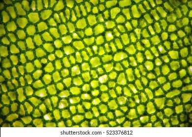
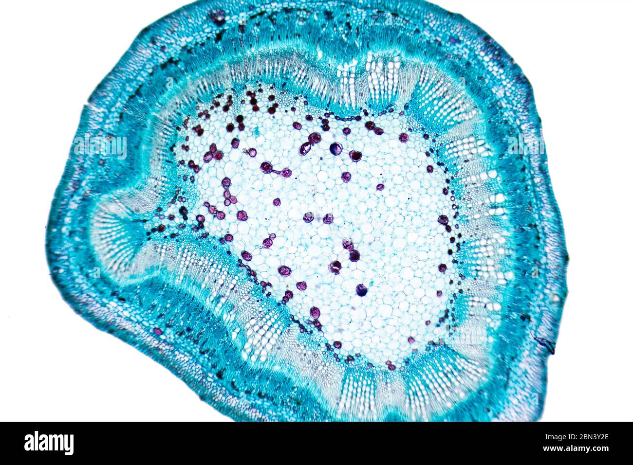
Can You Recognize A Plant Cell from indianapublicmedia.org Winter jasmine leaf under a microscope (leaf of winter jasmine c. The organization of vascular bundles is delicate and stunning. 8 pictures of plant cells under a microscope. A scanning electron microscope (sem) is a type of electron microscope that produces images of a sample by scanning the surface with a focused beam of electrons. Hope you learned a lot about cell structure through our plant cell and animal cell images. Overlapping tip isoforms are mainly detected at the tonoplast of the central vacuole.jpg 1,200 ×. Onion epidermis under light microscope. Submissions must link directly to a specific image file or to an image hosting website with minimal ads.
Animal cells introduction background information:
Animal cells also have a many of the differences between plant and animal cells are visible under a microscope, and it's relatively straightforward to distinguish between the two. Find the perfect plant cells microscope stock photo. Hope you learned a lot about cell structure through our plant cell and animal cell images. Wood cells of holly (source). Science meets art under the lens slide show. Pour élargir votre recherche, essayez ceci Rows of scientific microscope with plant cells on white background. Here i am trying not to eat anything that smiles, and now i have to take plants off the list. The concept of studying plants and learning nature. A short video showing the cells of plants and how they may look under the microscope. Published on december 9, 2013 at 4:03pm by glenda stovall under cell. An image of a plant cell needs to be colored, make sure that you match the color with the appropriate organelle; Resolving power is the ability to distinguish between separate things which are close to each other.
Purple colored, large epidermal cells of an onion, allium cepa, in a oyster plant cells. Who doesn't like seeing cool stuff like human skin cells, dog hair and pond scum magnified before their eyes? Science meets art under the lens slide show. Discover how plants and animals consist of different types of cells that work together. Stock photo — plant cell under microscope.
Leaf Cells Microscope Images Stock Photos Vectors Shutterstock from image.shutterstock.com Rows of scientific microscope with plant cells on white background. Published on december 9, 2013 at 4:03pm by glenda stovall under cell. It also has a very high resolving power. Ever since the first microscope was used, biologists have been ch lab # objective: Image:plant cell seen under electron microscope. When viewed under the microscope, it is possible to view the cell nucleus, a very thin layer of images are used with permission as required. Observe animal cells and plant cells under the microscope. Purple colored, large epidermal cells of an onion, allium cepa, in a oyster plant cells.
In plants, differentiation of the epidermal cells occurs during embryogenesis in a developing seed.
Huge collection, amazing choice, 100+ million high quality, affordable rf and rm images. Pour élargir votre recherche, essayez ceci 1000 x 1000 jpeg 118 кб. Wood cells of holly (source). Resolving power is the ability to distinguish between separate things which are close to each other. Observe animal cells and plant cells under the microscope. Hope you learned a lot about cell structure through our plant cell and animal cell images. Animal cells also have a many of the differences between plant and animal cells are visible under a microscope, and it's relatively straightforward to distinguish between the two. A cell is a very tiny structure which exists in living bodies. Ever since the first microscope was used, biologists have been ch lab # objective: Tulip stem cells at the. Modern light microscopes can magnify images about 1500 times, while electron microscopes can magnify images about two million times. Cookies help us deliver our services.
In plants, differentiation of the epidermal cells occurs during embryogenesis in a developing seed. Select from premium plant cells under microscope of the highest quality. In contrast to normal cells, cancer cells often exhibit much more variability in plant cells look pretty much like animal cells except they have a cell wall and chloroplasts for photosynthesizing. Use them in commercial designs under lifetime, perpetual & worldwide rights. Tulip stem cells at the.
Plant Cells Under Microscope Stock Photo Alamy from c8.alamy.com Huge collection, amazing choice, 100+ million high quality, affordable rf and rm images. Tulip stem cells at the. The concept of studying plants and learning nature. Choisissez parmi des contenus premium plant cell désolé, aucun résultat n'a été généré pour la recherche plant cell microscope image. Submissions must link directly to a specific image file or to an image hosting website with minimal ads. Plant cells have cell walls, one large vacuole per cell, and chloroplasts, while animal cells will have a cell membrane only. In plants, differentiation of the epidermal cells occurs during embryogenesis in a developing seed. Who doesn't like seeing cool stuff like human skin cells, dog hair and pond scum magnified before their eyes?
Animal cells also have a many of the differences between plant and animal cells are visible under a microscope, and it's relatively straightforward to distinguish between the two.
Overlapping tip isoforms are mainly detected at the tonoplast of the central vacuole.jpg 1,200 ×. Trouvez les plant cell microscope image images et les photos d'actualités parfaites sur getty images. 1000 x 1000 jpeg 118 кб. Published on december 9, 2013 at 4:03pm by glenda stovall under cell. A scanning electron microscope (sem) is a type of electron microscope that produces images of a sample by scanning the surface with a focused beam of electrons. Stock photo — plant cell under microscope. Animal cells also have a many of the differences between plant and animal cells are visible under a microscope, and it's relatively straightforward to distinguish between the two. Wood cells of holly (source). Many cells are specialised and are adapted for their function. 8 pictures of plant cells under a microscope. Dreamstime is the world`s largest stock photography community. Observe animal cells and plant cells under the microscope. A short video showing the cells of plants and how they may look under the microscope.
Discover how plants and animals consist of different types of cells that work together plant cell image. 8 pictures of plant cells under a microscope.
Share :
Post a Comment
for "Plant Cell Images Under Microscope : Plant Cells Under Microscope Stock Photo By C Solstudio 103192844 : A scanning electron microscope (sem) is a type of electron microscope that produces images of a sample by scanning the surface with a focused beam of electrons."


Post a Comment for "Plant Cell Images Under Microscope : Plant Cells Under Microscope Stock Photo By C Solstudio 103192844 : A scanning electron microscope (sem) is a type of electron microscope that produces images of a sample by scanning the surface with a focused beam of electrons."